When TMA Strikes - Every Second Counts
Early identification of the underlying cause of the TMA is critical for patient outcomes.
Patients with TMA present with the following clinical triad of laboratory signs and symptoms:
And one or more of the following:

Read McFarlane PA, et al. Making the Correct Diagnosis
in Thrombotic Microangiopathy: A Narrative Review.
TMA is a symptom, not a diagnosis. TMAs can share the same clinical presentations but differ in the underlying cause. Determining the origin is crucial for initiating appropriate treatment.2,3
Adapted from McFarlane PA, et al. Can J Kidney Health Dis. 2021;8:20543581211008707.
aHUS, atypical hemolytic uremic syndrome; CFH, complement factor H; DGKE, diacylglycerol kinase epsilon; DIC, disseminated intravascular coagulation; HSCT, hematopoietic stem cell transplantation; HUS, hemolytic uremic syndrome; INF2, inverted formin-2; STEC-HUS, Shiga toxin E. coli related hemolytic uremic syndrome; TMA, thrombotic microangiopathy; TTP, thrombotic thrombocytopenic purpura.
*TMA forms predominantly affecting children.
Urgently run key tests, including ADAMTS13 and Shiga Toxin panel, to differentiate between aHUS, STEC-HUS, and TTP3
- A PLASMIC score of 0 to 4 can be helpful to rule out TTP5
- A patient with TMA presenting a PU/CU of ≥1.5 g/g is less likely to have TTP6
- A platelet count >30 x 109/L and/or serum creatinine >1.7 to 2.3 mg/dL almost eliminates a diagnosis of severe ADAMTS13 deficiency (TTP)1
TTP, STEC-HUS, and aHUS are driven by different pathophysiologic processes and have different management goals
While there is no single, rapid, or definitive test to positively diagnose aHUS, it should be suspected in the presence of TMA3:
- With at least partially preserved ADAMTS13 activity and absence of a coexisting condition or drug exposure known to trigger TMA, or
- When the TMA fails to improve despite adequate treatment of a coexisting condition
- Shiga toxin negative test rules out STEC-HUS
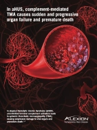
In aHUS, uncontrolled terminal-complement activation leads to systemic TMA, causing progressive damage to vital organs and premature death.2,3
Triggers are conditions that can activate complement and may unmask aHUS. It is imperative to treat the trigger, but if the signs and symptoms of TMA do not resolve, consider a diagnosis of unmasked aHUS.


Complement-amplifying conditions can help unmask aHUS
Recommendations for Suspected aHUS in the ICU2,*
- Obtain appropriate diagnostic samples before plasma therapy.
- Make use of multidisciplinary care, including nephrologists and hematologists.
- Initiate plasma therapy within 4-8 hours of admission or diagnosis of TMA.
- For patients with an initial presentation of TMA, switch to a complement inhibitor as soon as a diagnosis of aHUS is confirmed (ADAMTS13 > 10% and STEC negative);
- For patients with a history of previous aHUS, initiate complement-inhibitor therapy immediately on admission to the ICU.
- Careful ICU monitoring—organ dysfunction may appear or worsen until remission.
- Because of the increased risk of Neisseria meningitidis infection with complement-inhibitor treatment, patients should be vaccinated against serotypes A, C, Y, and W135 and subtype B two weeks before complement-inhibitor therapy is initiated; unvaccinated individuals should receive prophylactic antibiotics on complement-inhibitor therapy initiation until at least 2 weeks after Neisseria meningitidis vaccination.
*Adult patients with aHUS
Without proper treatment, aHUS has a high degree of morbidity and mortality7


PE/PI is not enough7
Outcomes for patients with aHUS receiving PE/PI are poor—up to 56% of adults progress to ESRD or die within 1 year8 and up to 77% have ESRD or die within 3 years9
- If TMA and aHUS are suspected, consult a multidisciplinary team of specialists including hematologists in the diagnostic process
- Follow the differential diagnosis pathway to reach a diagnosis
The treatment of choice for aHUS is complement inhibition3
Early clinical intervention reduces the risk of irreversible
organ injury and life-threatening complications1,2
organ injury and life-threatening complications1,2
ADAMTS13, a disintegrin and metalloproteinase with a thrombospondin type 1 motif, member 13; aHUS, atypical hemolytic uremic syndrome; ICU, intensive care unit; PE, plasma exchange; PI, plasma infusion; PU/CU, proteinuria/creatininuria ratio; TMA, thrombotic microangiopathy; STEC, Shiga toxin-producing E. coli; STEC-HUS, shiga toxin E. coli related hemolytic uremic syndrome; TTP, thrombotic thrombocytopenic purpura.